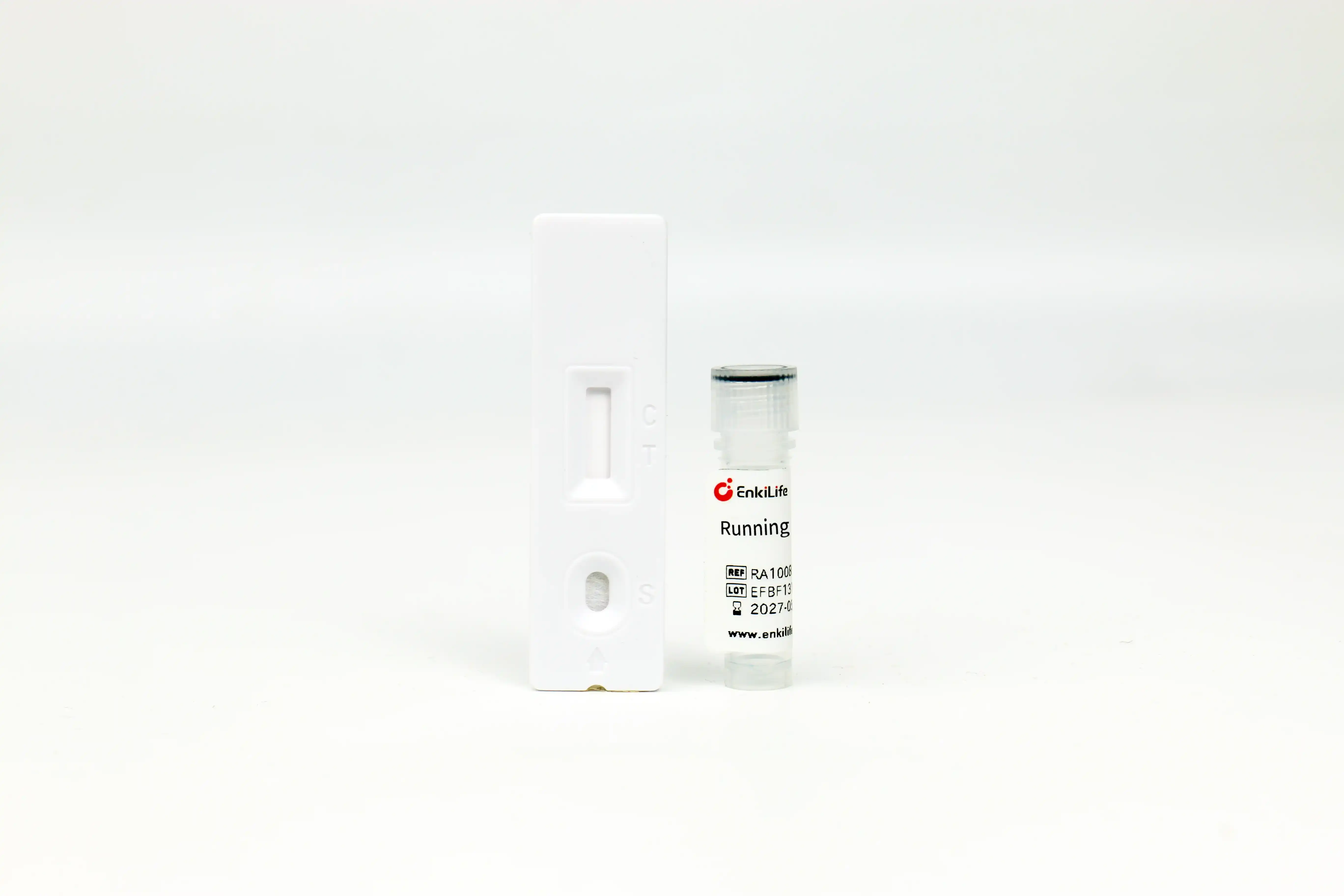



V5-Tag Rapid Competitive Assay Kit
Dedicated to rapid detection of V5-tagged Protein · Results available in 10-15 min · High specificity based on competition principle
Product Overview
V5-Tag Rapid Competitive Assay Kit adopts colloidal gold lateral flow chromatography technology and is developed based on competitive immunoassay principle. Recombinant V5-Tag protein is immobilized on the chromatographic membrane as test line, and secondary antibody for colloidal gold conjugated antibody is fixed as control line; colloidal gold-labeled anti-V5-Tag monoclonal antibody is pre-coated on the conjugate pad.
If no V5-Tag protein exists in samples, gold-labeled antibodies migrate freely to the test line and bind with immobilized V5-Tag protein to form distinct purplish-red bands. When V5-Tag target protein is contained in samples, the target binds preferentially to gold-labeled antibodies and occupies antigen-binding sites, blocking the combination between gold-conjugated antibodies and immobilized protein on test line. Higher concentration of V5-Tag protein leads to lighter color of test line; full disappearance of test line occurs above 18 μg/mL protein concentration. The limit of detection is 2 μg/mL, and test line color intensity is negatively correlated with protein concentration within 2 μg/mL ~ 18 μg/mL.
Core Advantages
Time Comparison with Conventional Methods
This kit shortens total protein detection duration from a full day to merely 15 minutes!
Product Specifications
| Catalog No. | Pack Size | Storage Condition | Shelf Life |
|---|---|---|---|
| RA10087 | 10T | Dry ambient temperature | 12 months |
| RA10087 | 100T | Dry ambient temperature | 12 months |
Kit Components: Individually foil-sealed test cassettes, dropper bottle of running buffer and detailed product manual
Wide Application Fields
- Screening of recombinant protein-expressing clones: Rapid identification of high-expression positive transformants to avoid unnecessary consumption of reagents and incubation on low-yield strains
- Optimization of protein expression conditions: Fast comparison of expression levels under varied induction temperature, duration and inducer concentration
- Tracking throughout protein purification: Real-time quantification of target protein in loading sample, flow-through and elution fractions
- Transfection efficiency detection for eukaryotic cells: Quick evaluation of target protein expression from different transfection reagents and protocols
- Qualification verification of protein samples: Confirm integrity of affinity tag before downstream functional assays
- Mass production quality control: Intermediate batch testing for recombinant biopharmaceuticals and industrial enzyme production
Operating Procedure
- Preparation: Take detection cassette out from sealed aluminum pouch and place horizontally on dry bench; dilute sample appropriately to adjust target protein concentration within optimal testing range of 2–18 μg/mL
- Sample Loading: Pipette 20 μL diluted specimen slowly into sample well of test cassette
- Initiate Chromatography: Immediately add 50 μL matched running buffer (approx. 2 drops vertically from dropper) into sample well
- Result Reading: Incubate at room temperature for 10–15 min before interpretation; readings taken after 20 min are invalid.
Sample Dilution Guide: For samples with known concentration, dilute to ~6 μg/mL directly; for unknown bacterial lysate, start with 20-fold dilution; for mammalian cell supernatant or lysate, perform initial 4-fold dilution. If faint or absent test line appears with expected high protein expression, further dilute sample by 10 folds and retest.
Guidance for low-abundance protein: Undiluted raw sample can be used directly for samples with extremely low target expression; always run matching matrix negative control to differentiate band intensity for valid judgement.
Result Interpretation
Both control line (C) and test line (T) develop purplish-red bands with identical color intensity to negative control, indicating no V5-Tag protein or target concentration below 2 μg/mL LOD in specimen.
Control line (C) is visible, while test line (T) is completely absent or markedly lighter than negative control, confirming presence of V5-Tag protein; paler test line corresponds to higher protein concentration.
No visible band at control line (C) regardless of test line status; invalid testing caused by expired reagent, improper operation or insufficient running buffer volume.
Compatibility with Common Lab Reagents
The kit tolerates multiple routine buffer additives, enabling direct testing of most routinely prepared protein lysates without extra pre-treatment.
| Reagent Name | Max Tolerated Concentration |
|---|---|
| NaCl | 1.5M |
| KCl | 1.5M |
| Urea | 0.4M |
| Triton X-100 | 1% |
| Tween-20 | 1% |
| Reagent Name | Max Tolerated Concentration |
|---|---|
| NP-40 | 1% |
| CHAPS | 1.0% |
| SDS | 0.20% |
| EDTA | 7.5mM |
| Glycerol | 10% |
Special Note: Fully compatible with undiluted RIPA lysis buffer; cell lysates prepared in RIPA can be tested directly without dialysis or further processing.
Precautions
- Intended solely for academic research use; prohibited for human clinical diagnosis or therapeutic application.
- Test cassette must be used within 1 hour after foil pouch opening; prolonged ambient exposure leads to moisture-induced reagent deactivation.
- Appropriate sample dilution is critical to reliable results; adjust dilution fold based on anticipated target concentration.
- Negative control is mandatory for competition-based assay to judge band intensity variation accurately.
- Unexpected pale or missing test line against expected expression level suggests over-high analyte concentration; dilute sample further and repeat assay.
- Strictly follow stipulated incubation time for result reading; premature or delayed interpretation leads to false conclusion.
- Do not mix test cassettes and running buffer from different production lots as minor batch-to-batch performance variation exists.
- Dispose all experimental waste in accordance with institutional biosafety regulations.
Troubleshooting Guide
| Observation | Possible Causes | Solutions |
|---|---|---|
| No obvious fading of test line vs negative control | Absence of target protein Protein concentration below LOD |
Verify matching of expression vector and test kit Retest with undiluted raw sample plus negative control |
| Extremely faint or fully absent test line | Sample concentration exceeds upper detection limit Complete competitive inhibition from over-high antigen level |
Dilute sample at higher fold and perform repeated testing |
| No band formed at control line | Incorrect operating steps Expired test strips Improper humid storage of cassettes Insufficient running buffer added |
Repeat test following full manual instruction Replace with unexpired new test cassette Store products under cool & dry environment Add adequate running buffer (~2 drops) |